Nanoscale and Condensed
Matter Physics
The fundamental understanding of the electronic, structural, chemical
and optical properties of materials on the nanoscale is essential
for advances in nanotechnology. Developments in experimental physics
underpin these advances via characterisation and quantification of
quantum phenomena which can dominate at these length scales. Work
at Swansea under the direction of Peter Dunstan has concentrated particularly
on the optical properties of materials on the nanoscale. Using advances
in scanning probe microscopy, and in particular scanning near-field
optical microscopy, semiconductor materials/devices and soft organic
materials have been the subject of investigation.
The group is an active member of the University's Multidisciplinary Nanotechnology Centre (MNC), with our laboratories forming part of the central facility on campus. Also see detailed research link on MNC webpages.
Current research team
Omar Al-Hartomy (writing up PhD)
Kelly-Ann Walker (3rd year PhD)
Jonathan Lloyd (2ndd year PhD)
Former students
Gareth John (PhD 2005)
Mathew Ackland (PhD 2005)
Jeff Kettle (PhD 2007)
Mark Holton (PhD 2007, Post-doc within MNC)
Richard Baylis (PhD 2007)
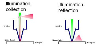
Nano-optics and scanning near-field optical microscopy
Imaging materials with visible light has traditionally had its limits, namely optical diffraction. To enable nanoscale optical imaging a specific sample-light source arrangement is required which overcomes the limitations of far-field light and exploits the near-field regime and its non-propagating evanescent components. This can be achieved using the technique of scanning near-field optical microscopy (SNOM or NSOM) which usually employs a tapered optical fibre with a nano-sized (50 -100 nm) aperture at the end. Light is spatially confined by this arrangement and thus it can be used as a nanoscale source of light. To ensure that the non-propagating evanescent field is utilised the tip-sample distance must be controlled to within several nanometres using scanning probe microscopy. This microscopy technique can be employed in a number of configurations, as shown below:

Current areas of research and examples of recent results
EPSRC funded research into colloidal quantum dots probed by near-field excitation: These Q-dots have a tuneable fluorescence depending on their size and thus our current activities are immobilising the quantum dots in a polymer film to probe their absorption and emission properties. Significant developments are being made to our instrumentation to enable high-resolution spectroscopy on single quantum dots.
The MNC funds a joint PhD studentship project with Cardiff University
to consider the role of surface plasmons in light emission from nanostructured
surfaces. The regular lattice (or grating) of apertures generated
in a metal film are being studied, particularly the role the near-field
has in the resulting enhanced optical emissions.
Examples
1. Collection mode from vertical cavity surface emitting lasers (VCSEL)
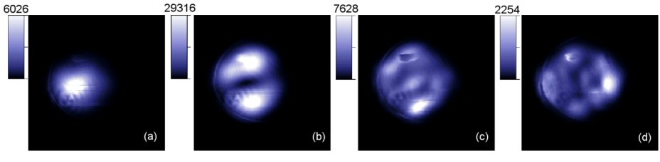
Narrow-bandwidth, wavelength specific, near-field optical collection images (20µm x 20µm) from the surface of the VCSEL run at 2.34mA.
Wavelengths are (a) 845.56nm, (b) 845.37nm, (c) 845.19nm and (d) 844.95nm.
2. Photocurrent measurements made from a buried heterostructure QW laser

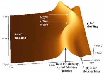
Topographic and photocurrent images taken from the active region of a buried hererostructure QW laser.
3. Near-field imaging of tagged human chromosomes


Topographic and fluorescent images of human chromosome 1 labelled with FITC whole chromosome paint with signal amplification (12.4µm x 12.4µm with 3µm size bar).